Data analysis
Hyperion allows for single-cell, spatially resolved, highly multiplexed (>40 parameters) analysis of solid tissues. To provide a complete picture of a tissue ecosystem, define molecular and spatial signatures, it is necessary to analyze and interrelate information obtained from molecular measurements on cells, cell-cell interactions, and microenvironments. There are several options available to analyze high dimensional imaging mass cytometry data.
We recommend getting in touch with a data analyst already in the planning phase of your experiments to ensure that the data you generate is of sufficient quality and all necessary controls are measured. At present, the IMC platform is not able to provide the service of data analysis but will assist you in finding a solution.
Preliminary Hyperion data analysis
This will provide you with the cell subsets present and perform a basic neighborhood analysis. For comparison of conditions and statistics, further data analysis is required by a designated bioinformatician.
Quality control
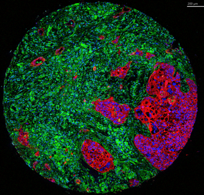
MCD™ Viewer is post-acquisition data processing software that allows users to visualize, review, and export Imaging Mass Cytometry™ data acquired with the Hyperion™ Imaging System and CyTOF® Software v7.0
MCD Viewer Software (https://www.fluidigm.com/software). This can be done before cell segmentation to determine the quality of your staining.
The 6 main steps of imaging mass cytometry (IMC) basic analysis are as follows
- Preprocessing and filtering
- Pixel classification
- Image segmentation and quantification of single cells
- Single-cell and neighbourhood analysis
- Dimensionality reduction
- Clustering
Protocol for IMC basic analysis covering steps 1-3 above
Here is an SOP for obtaining single-cell data from raw IMC data, which covers steps 1,2 and 3 of data analysis. It is based on Vito Zanotelli`s pipeline.
Vito RT Zanotelli, Bernd Bodenmiller, ImcSegmentationPipeline: A pixel classification based multiplexed image segmentation pipeline (https://github.com/BodenmillerGroup/ImcSegmentationPipeline)
It performs image preprocessing, uses ilastik for supervised pixel classification, and CellProfiler for image segmentation and quantification of single cells. It provides users with cell mask(s) and TIFF files that can be used in histoCAT, where both single cell and neighborhood analysis can be performed or extracts the single-cell target expression and position information. Such extracted data from the image can be used for further high dimensional data analysis in FlowJo/Cytobank/any programming language.
IMC segmentation pipeline documentation
Slides from Bodenmiller workshop on cell segmentation
Protocol for IMC basic analysis covering steps 4-6 above
Histology Topography Cytometry Analysis Toolbox (histoCAT) is a package to visualize and analyse multiplexed image cytometry data interactively (https://www.nature.com/articles/nmeth.4391).
Corresponding manual and downloads are available (histoCAT).
With this analysis tool, users can make perform dimensionality reduction (tSNE), clustering (Phenograph) and neighborhood analysis to identify their cell types and their microenvironment.
Cytomapper is a R/Bioconductor package contains functions for the visualisation of multiplexed read-outs and cell-level information obtained by multiplexed imaging technologies. The main functions of this package allow the visualisation of pixel-level information across multiple channels, the display of cell-level information (expression and/or metadata) on segmentation masks and gating and visualisation of single cells.
All analysis code and instructions for data analysis are available (cytomapper)
The release version of the cytomapper package (version 1.0.0) can be installed via Bioconductor (https://www.bioconductor.org/packages/release/bioc/html/cytomapper.html)