Hyperion
Hyperion Imaging Mass Cytometry (IMC) is based on laser ablation technology coupled with time-of-flight (TOF) mass cytometry. This technology allows for a comprehensive understanding of complex cellular phenotypes and their interrelationships in the spatial context of the tissue microenvironment. In contrast to conventional immunohistochemistry or immunofluorescence, IMC is capable of the simultaneous analysis of >40 specific protein markers on tissues at subcellular resolution with minimal spectral overlap and low autofluorescence
Imaging Mass cytometry pipeline
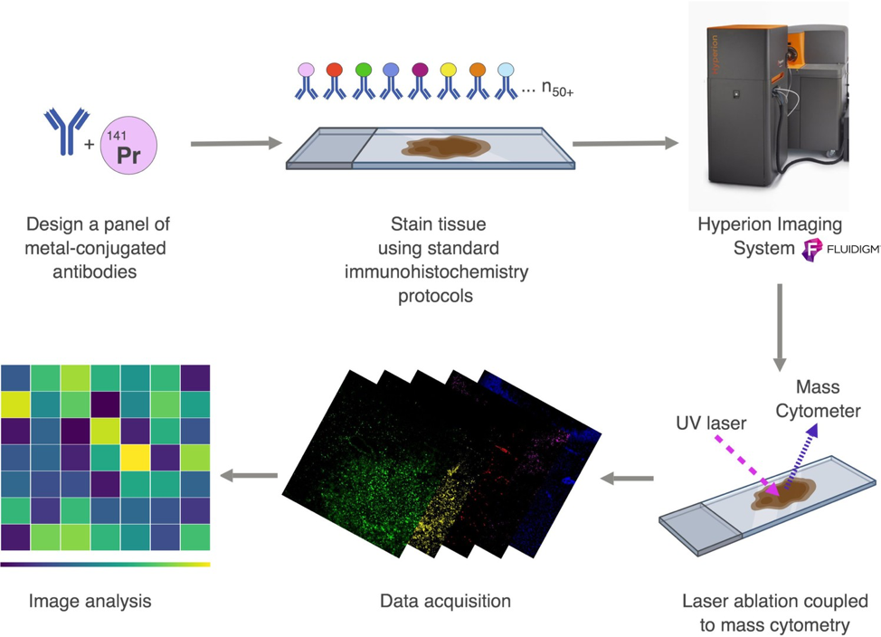
The Hyperion Imaging System consists of a Helios instrument, with an imaging module attached to the front. Tissue sections are first labelled with metal-isotope- conjugated antibodies/probes. Within the imaging module, the tissue sections are ablated by a high power pulsed UV laser with 1µm spot size. The resulting plumes of ablated tissue atoms are then transported to Helios, where the arriving atoms are ionized, and the metal ion abundances are measured by TOF mass spectrometry. The metal isotope abundance of each spot (in pixels) are then mapped back to the original co-ordinates, generating a high-dimensional image, which can be analysed using any third party software.
An example of publications using the Hyperion can be found here